Enjoy Disect Pro, completely Free.

The Disect Company was originally started in the early 2000’s by three individuals: Dr Duncan Bell, a consultant physician with an interest in imaging systems , Professor David Heatley, a telecommunication expert based on BT’s Martlesham Research site and thirdly an extremely talented software engineer, the late Dr Roger Rowland.
Disect Pro gradually evolved into the Company’s premium imaging software solution thanks to the advice and help of a team of Radiologists, Consultant surgeons, Anatomists and Educational experts based at the Ipswich Hospital and the Medical School at UEA, Norwich. Later, with the involvement of Dr Mark Greco, an Australian Entomologist with a background in Radiography and clinical CT, the software was slightly adapted to cater for micro-CT images in general and those from insect specimens in particular and marketed as ‘Bee View’.
It is an unfortunate fact that good ideas and technology do not always
translate into commercial success and the Disect Company was forced to stop trading some years back. Despite this, Professor Heatley has continued to make the Disect Pro software freely available to a small number of research workers without charge. We feel that Its powerful 3D visualization capabilities with user-friendly features, make it ideal for schools, FE colleges, medical or veterinary education, and for various research applications.
With this aim in mind, Duncan decided to start a YouTube portal called ‘Beeview’ to show case some of the applications of Disect Pro. He was ably helped in this venture by Mr George Balding, a UEA Media Studies graduate. Simultaneously Duncan was aided by both Mr Harri Mortimore and Dr David Mortimore, the author of the ‘Tomomask’ software, in opening the present website ‘Disect Revisited’.


The website permits the free download of the Disect Pro software and
many fascinating image files that can be read into the software. The software ‘reads’ DICOM clinical files as well as files in the form of its own file format( DsVs) and also transverse image stacks of TIFFs, BMPs etc. This makes it ideal for looking at micro-CT images which are not normally available as DICOM files. There are an increasing number of repositories for such files available around the World. By making Disect Pro freely available at no cost, we hope that individuals, schools and FE colleges all over the World can still have the excitement and educational benefit of viewing a wide variety of fascinating 3-D image files.
Summary of Features:
EASE OF USE
The method of loading data from all sources is straight forward and all image manipulation functions are available in a number of different ways. These include standard menu options, toolbar buttons, right-click context menus, slider and combo-box controls on the control panel, and dedicated mouse functions. In presentation view, where the toolbar, status bar and control panel are hidden, all of the common image manipulation functions can be performed using a conventional mouse or track pad.
simultaneous 2d & 3d views
Disect Pro can optionally display a split screen containing four views of the current DICOM data – a 3D reconstruction, the original 2D tomographic images (usually axial), and two reconstructed 2D orthogonal projections. For the 3D view, this is only relevant when the Multi-Planar Reconstruction (MPR) view is selected. The display may be toggled between the four-way split and full-screen view of any of the windows simply by clicking on the appropriate icon on the menu toolbar.
interactive window level
Image data sets from scanners typically include more information than can be shown on screen. For example individual image points in the data may be stored using 4096 intensity levels for 12bit data and 65536 levels for 16bit data, but they need only be represented on screen in 256 levels of grey (unless the image is colourised using the display LUT). All medical imaging workstations provide a way for the source data to be mapped to the display known as windowing.
zooming
Any of the views in Disect Pro may be zoomed in or out simply using a click-drag with the mouse once the zoom option has been selected.
Optionally zoom control for the 3D may use the control wheel on the lower edge of the viewing area. Zoomed images may also be saved as screenshots or copied to the clipboard for pasting into a 3rd party application.
2d view features
Click-drag to pan around a zoomed image, scroll through consecutive slices, drag a cursor to roam around an individual slice while synchronising the other views, or flip an image left/right or up/down to take account of different patient orientations during the original scan. A measurement tool allows linear measurements to be made from any of the 2D views (CT data only).
3d view features
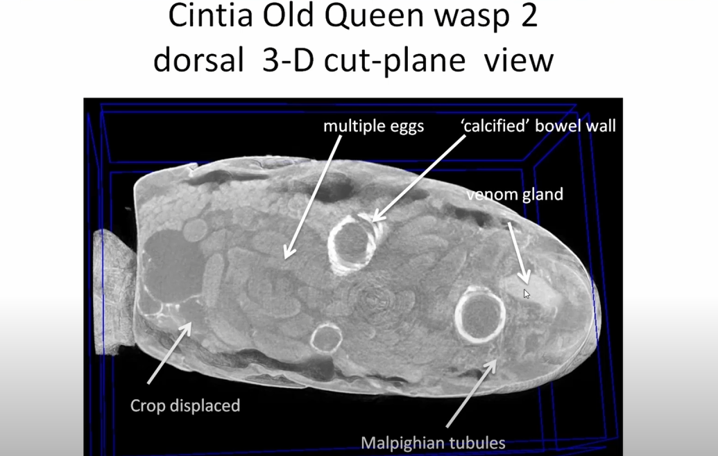
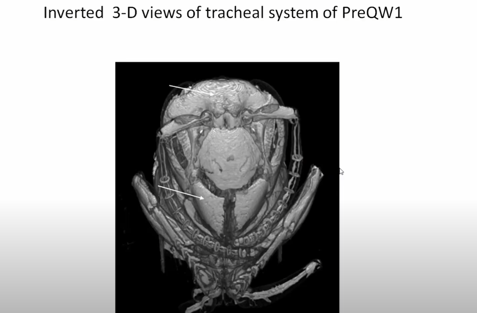
The 3D reconstructed views produced by Disect Pro include a 3D volume view with up to 6 moveable orthogonal cut-planes, a maximum intensity projection (MIP) view, and a multi-planar reconstruction (MPR) view.
The MPR view is automatically synchronised with scroll and roam actions from any of the 2D views.
Download Disect Pro Now
Disclaimer
The user accepts the Software “as is” and The Supplier makes no warranty as to its use, performance, or otherwise. To the maximum extent permitted by applicable law, The Supplier disclaims all other representations, warranties, and conditions, express, implied, statutory, or otherwise, including, but not limited to, implied warranties or conditions of merchantability, satisfactory quality, fitness for a particular purpose, title, and non-infringement.
The permitted use of the Software is for visualisation and education purposes only.
Further details are contained in the End User License Agreement for the software.